lv aneurysm ecg | left ventricular pseudoaneurysm vs aneurysm lv aneurysm ecg A left ventricular aneurysm can be diagnosed on ECG when there is persistent ST segment elevation occurring 6 weeks after a known transmural myocardial infarction (usually an anterior . In patients with ischemic LV systolic dysfunction, CABG plus medical therapy resulted in higher mortality at 30 days, but with a significant improvement in long-term mortality (out to 10 years) compared with medical therapy alone. Only 14 patients needed to be treated with CABG to save one life over 10 years.
0 · what is ventricular aneurysm
1 · what is an apical aneurysm
2 · ventricular aneurysm ecg
3 · left ventricular pseudoaneurysm vs aneurysm
4 · left ventricular aneurysm repair surgery
5 · Lv pseudoaneurysm vs true aneurysm
6 · Lv aneurysm vs pseudoaneurysm echo
7 · Lv aneurysm on echo
GPSPRO.LV . Jaunmoku 26 (pie Komforta) blakus t/c Spice 20 015 015, par pasūtījumu 27 800 684. [email protected]. Informācija; Kontaktinformācija; Par mūsu uzņēmumu; Preču piegādes noteikumi; Jauni produkti; Produkti ar atlaidēm; Atgriešanās politika; Rīki; Iepirkumu grozs;
Learn how to identify and differentiate left ventricular aneurysm (LVA) from acute STEMI on the ECG. See examples, pathophysiology, clinical significance and references for LVA diagnosis.A left ventricular aneurysm can be diagnosed on ECG when there is persistent ST segment elevation occurring 6 weeks after a known transmural myocardial infarction (usually an anterior . A significant left ventricular (LV) aneurysm is present in 30% to 35% of acute transmural myocardial infarction. The two major risk factors for .Learn about ECG changes in STEMI, left ventricular (LV) aneurysm, and ST elevation dynamics. Explore T/QRS ratio (V1-4) and its clinical significance.
what is ventricular aneurysm
what is an apical aneurysm
The electrocardiographic features of LV aneurysm include the following: 1) STE, most often less than 3-4 mm; 2) diminished or inverted T waves; 3) QS or Qr complexes preceding the STE in the right-to-mid .ECG. 1. 30min - hours. Hyperacute T waves. >6mm limb leads. >10mm precordial leads. Normalizes in days, weeks, or months. 2. Minutes - hours.
Left ventricular aneurysm formation following acute STEMI causes persistent ST elevation on the ECG. ECG Features of Left Ventricular Aneurysm. ST elevation seen > 2 weeks following an acute myocardial infarction. Most commonly seen in the precordial leads. May exhibit concave or convex morphology. Usually associated with well-formed Q- or QS waves
A left ventricular aneurysm can be diagnosed on ECG when there is persistent ST segment elevation occurring 6 weeks after a known transmural myocardial infarction (usually an anterior MI)..
A significant left ventricular (LV) aneurysm is present in 30% to 35% of acute transmural myocardial infarction. The two major risk factors for developing LV aneurysm include total occlusion of the left anterior descending artery .Learn about ECG changes in STEMI, left ventricular (LV) aneurysm, and ST elevation dynamics. Explore T/QRS ratio (V1-4) and its clinical significance. The electrocardiographic features of LV aneurysm include the following: 1) STE, most often less than 3-4 mm; 2) diminished or inverted T waves; 3) QS or Qr complexes preceding the STE in the right-to-mid precordial leads; and 4) lack of dynamic change of these findings over time on ECG.
ECG. 1. 30min - hours. Hyperacute T waves. >6mm limb leads. >10mm precordial leads. Normalizes in days, weeks, or months. 2. Minutes - hours.
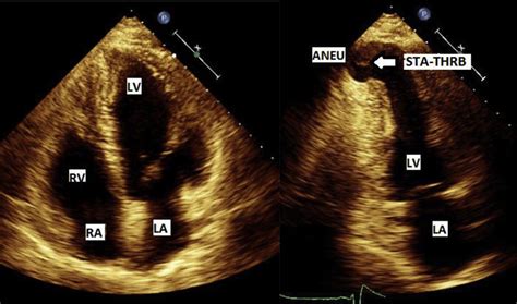
Persistent ST elevation after a STEMI can signify a left ventricular (LV) aneurysm. Differentiating LV aneurysm from STEMI is very challenging, as patients with an LV aneurysms are at high risk for cardiac pathology. If available, the crux of management is comparing the current ECG with an old ECG, which may show the persistent LV aneurysm pattern.The usual ECG findings of left ventricular aneurysm include ST elevation that persists more than two weeks after STEMI, deep Q waves, and the absence of reciprocal ST depressions. Apical four chamber echocardiogram showing severe balloon-like dilation of .ECG Findings: 1. Normal Sinus Rhythm. 2. Old Anterior Wall Myocardial Infarction. 3. Left Ventricular Aneurysm. LV aneurysms are often clinically silent and diagnosed on routine imaging. Otherwise patients can present with symptoms of heart failure, embolic event, or ventricular arrhythmia. Persistent ST-elevation on 12 lead-ECG may be seen, but has a low sensitivity and specificity for the presence of aneurysm.
Left ventricular aneurysm formation following acute STEMI causes persistent ST elevation on the ECG. ECG Features of Left Ventricular Aneurysm. ST elevation seen > 2 weeks following an acute myocardial infarction. Most commonly seen in the precordial leads. May exhibit concave or convex morphology. Usually associated with well-formed Q- or QS wavesA left ventricular aneurysm can be diagnosed on ECG when there is persistent ST segment elevation occurring 6 weeks after a known transmural myocardial infarction (usually an anterior MI).. A significant left ventricular (LV) aneurysm is present in 30% to 35% of acute transmural myocardial infarction. The two major risk factors for developing LV aneurysm include total occlusion of the left anterior descending artery .
ventricular aneurysm ecg
Learn about ECG changes in STEMI, left ventricular (LV) aneurysm, and ST elevation dynamics. Explore T/QRS ratio (V1-4) and its clinical significance.
The electrocardiographic features of LV aneurysm include the following: 1) STE, most often less than 3-4 mm; 2) diminished or inverted T waves; 3) QS or Qr complexes preceding the STE in the right-to-mid precordial leads; and 4) lack of dynamic change of these findings over time on ECG.ECG. 1. 30min - hours. Hyperacute T waves. >6mm limb leads. >10mm precordial leads. Normalizes in days, weeks, or months. 2. Minutes - hours.
Persistent ST elevation after a STEMI can signify a left ventricular (LV) aneurysm. Differentiating LV aneurysm from STEMI is very challenging, as patients with an LV aneurysms are at high risk for cardiac pathology. If available, the crux of management is comparing the current ECG with an old ECG, which may show the persistent LV aneurysm pattern.
The usual ECG findings of left ventricular aneurysm include ST elevation that persists more than two weeks after STEMI, deep Q waves, and the absence of reciprocal ST depressions. Apical four chamber echocardiogram showing severe balloon-like dilation of .ECG Findings: 1. Normal Sinus Rhythm. 2. Old Anterior Wall Myocardial Infarction. 3. Left Ventricular Aneurysm.

Goodwill Rainbow Store. 741 S Rainbow Blvd. Las Vegas, NV 89145. 702-214-2028. Store & Donation Center Hours. Mon–Sat | 9am–9pm. Sunday | 9am–7pm.
lv aneurysm ecg|left ventricular pseudoaneurysm vs aneurysm


